Posts

Showing posts from October, 2018

Are SGLT2 inhibitors renoprotective?

Image
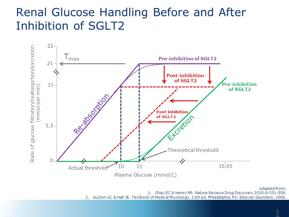
SGLT2 Inhibitors and Renoprotection Q: “How do SGLT2 inhibitors help in chronic kidney disease?” A: The renoprotective effect of SGLT2 inhibitors is still being extensively studied, but the results have been very promising. Let’s start with discussing what SGLT2 inhibitors are. SGLT 2 inhibitors SGLT2 inhibitors (eg dapagliflozin, canagliflozin, empagliflozin etc) are a class of drugs that inhibit reabsorption of glucose in the kidney (by inhibiting the sodium-glucose transport protein 2 - ie, SGLT2) and therefore lower blood sugar. Because of their use in lowering blood glucose levels, they are regularly used in the management of type 2 diabetes mellitus (and increasingly in type 1 diabetes as well). In addition to glycemic control, they have been shown to provide significant cardiovascular benefit in patients with type 2 diabetes. Mechanism Sodium Glucose cotransporters (SGLTs) are proteins that are found in different tissues that play an important role in mai

Taking an arrow to the (Kid)ney.

Image
Q: “Definitive diagnosis for diseases such as Wegener's and Churg Strauss is by renal biopsy. Is there some imaging done to determine where to do the biopsy from or are the granulomas so widespread that any part of the kidney can be biopsied?” A: So, the simple answer to whether imaging is done in Wegener's/Churg Strauss to determine a specific location in the kidney from where a biopsy is done is no! Points to keep in mind: Granulomatosis with polyangiitis (formerly known as Wegener’s) affects the kidney in approximately 50 % of the cases and present as focal and segmental necrotising granulomas in the kidney. These findings cannot be seen on imaging (CT, MRI) in most cases. Rarely (and I mean very rarely), they present as solitary/ multiple renal masses. Eosinophilic Granulomatosis with polyangiitis (formerly Churg-Strauss) affects the kidney in approximately 25% of the cases and presentation is same as that seen in Wegener’s and imaging does not help in ident

The IV League

Image
Q: “What is the difference between IV bolus, IV infusion, IV injection, IV push and IV drip?” Let’s look at what each of these terms mean: Intravenous injection : a hypodermic injection into a vein for the purpose of instilling a single dose of medication, injecting a contrast medium, or beginning an IV infusion of blood, medication, or a fluid solution, such as saline or dextrose in water. IV bolus : concentrated medication and/or solution given rapidly over a short period of time Eg.  Fluid bolus: a rapid administration of fluids to correct hypotensive shock. It typically includes the infusion of at least 500 ml over a maximum of 15 min. IV push : manual administration of medication under pressure, can be over several minutes; does not necessarily mean  in seconds. Most medications are given over more than a minute. ( IV push = IV bolus; the time over which a medication is given and its dilution, if any, varies) IV infusions: Continuous infusion – a lar

The method with Methotrexate.

Image
Methotrexate in Rheumatoid Arthritis Q: “At what stage of disease is methotrexate started in rheumatoid arthritis? Are there any criteria to start it?” A: I shall begin answering this question with talking about the drug methotrexate. Methotrexate Methotrexate is a chemotherapy agent and immune system suppressant. It is used to treat various types of cancer, autoimmune diseases, ectopic pregnancy and for medical abortion. In the context of its use in rheumatoid arthritis, it is classified as a DMARD (disease modifying antirheumatic drug). It works by inhibiting cytokinin production and purine biosynthesis with the inhibition of dihydrofolate reductase, and by stimulating adenosine release. The purpose of using methotrexate in rheumatoid arthritis is not for symptomatic relief. The goals of treatment are as follows: To control disease activity and joint pain To maintain the ability to function in daily activities To slow destructive joint changes To delay

Amniotic Abomination

Image
Q: How does amniotic fluid embolism cause Pulmonary Edema? A: To answer your question, we must take a peek into the pathophysiology of amniotic fluid embolism. It is a condition that can be characterised by the presence of Cardiovascular collapse : hypotension/cardiac arrest Respiratory distress : acute hypoxia Coagulopathy All of these occurring during labour, caesarean delivery, dilation & evacuation, or within 30 minutes postpartum with no other explanation of findings. [1] [5] In terms of pathophysiology, there is a lot that remains to be understood about amniotic fluid embolism. The various proposed theories for amniotic fluid embolism can be classified into two types: The classical theory that states that all the features of this disease are attributable to a mechanical obstruction of pulmonary arteries by debris comprising of amniotic fluid and fetal cells. [1]    2. Later theories which state that the manifestations are not due to physical o

1 Step at a Time

Image
Q: “How does one prepare for USMLE Step 1? Could you suggest us briefly how many months one should read First Aid and how many months does one take to finish UWorld?” A: At the outset, I'd like to say that the following answer is one that has been subjectively answered. The basis for the following points has been taken from shared experiences of the team at Dumb-CQs. We do not bear any affiliations with the resources we mention in this article. To answer your question, I would like to begin by discussing the anatomy of the Step 1 Exam. The Step 1 exam is an 8 hour exam comprising of 7 blocks of 60 minutes duration of around 40 questions each. In this exam you will be tested in the basic sciences viz. Anatomy, Biochemistry, Physiology, Pathology, Microbiology, Pharmacology, Behavorial Sciences, Ethics and Biostatistics. That being said, most of the questions are based on clinical encounters and patient profiles, so a solid understanding of disease presentations and

Dexametha-Zone

Image
Q: What is the importance of Low Dose and High Dose Dexamethasone Suppression Tests? A: Let's begin with the HPA (Hypothalamus-Pituitary-Adrenal) axis: Cortisol suppresses the release of both CRH from the hypothalamus as well as ACTH from the pituitary. Low Dose Dexamethasone Test: The Overnight DST which is a type of Low Dose DST is used to rule out a hypercortisolism state or the diagnosis of Cushing’s syndrome. It is based on the principle that in a normal individual a low dose of dexamethasone is sufficient to suppress the ACTH production and hence the cortisol levels. Procedure: Overnight Dexamethasone test : 1 mg of Dexamethasone is given at 11pm and the cortisol level is measured at 8am. Failure to suppress the cortisol level to less than 50 nmol/L signifies Cushing's syndrome. LDDST : 0.5mg Dexamethasone q6h for 2 days and then the plasma cortisol is measured. Plasma cortisol more than 50 nmol/L is considered abnormal. Any abnormalit