Cherry on the Top
What is a Cherry Red Spot?
A: To answer this we must start with the normal anatomy of the Macula:
Macula Lutea (commonly called the yellow spot) is a circular area located at the centre of the posterior retina, 2 disc diameters from the optic disc.
It has a slightly deeper red color when compared to the rest of the retina.
The fovea is the central 1.5mm of the macula and has a higher density of cones and is responsible for color vision. A central depression in the fovea known as the foveola is completely devoid of ganglion cells and rods and has the highest density of cones and hence the most acute vision.It is also relatively thinner when compared to the rest of the retina.
The Fovea receives its blood supply from the choroidal circulation via the long and short posterior ciliary arteries and NOT by the central retinal artery.
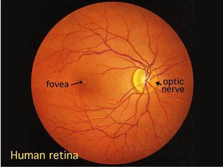 |
| Normal Retina |
Cherry Red Spot:
A Cherry Red spot is a fundoscopic finding at the Macula in various diseases. It is basically a bright or dull red spot surrounded by a greyish white halo.
It is commonly seen in the following diseases:
- Central Retinal artery Occlusion
- Macular hemorrhage
- Lysosomal Storage Diseases
- Tay-Sachs
- Neimann Picks disease
- Sandhoff’s disease
- Mucopolysaccharidosis
- Drug Toxicities
- Quinine
- Dapsone
- Methanol and Carbon monoxide poisoning
In CRAO and orbital ischemia:
As the blood supply of the fovea is different from that of the rest of the retina any embolus or occlusion of the central retinal artery leads to appearance of a pale infarcted surrounding retina with a bright red spot at the fovea.
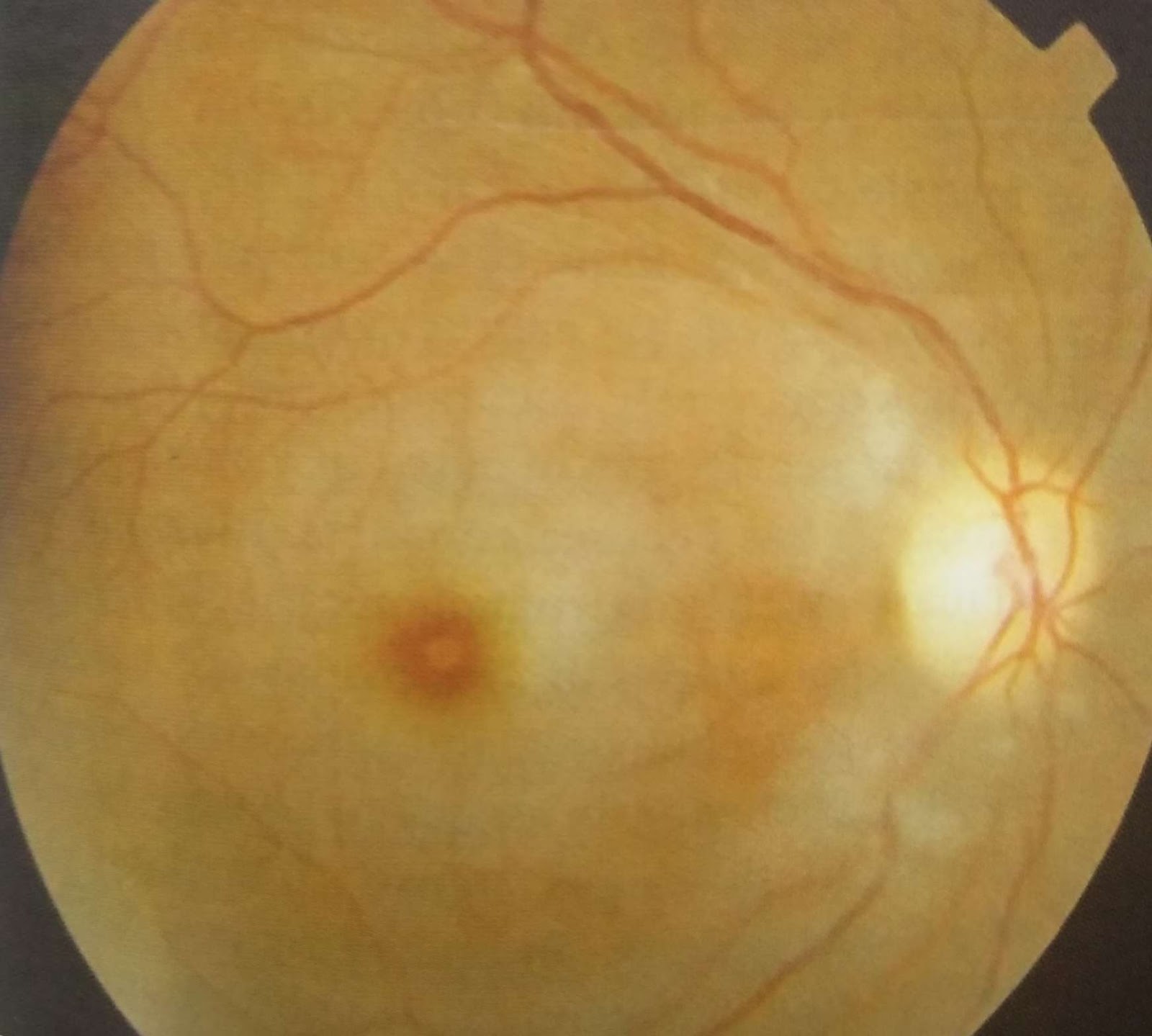
Cherry red spot in CRAO
In Lysosomal Storage Diseases:
The fovea of the macula is relatively thinner and completely devoid of ganglion cells. It is in these ganglion cells that the abnormal products of enzyme deficiencies (Gm2 ganglioside in Tay-sachs and Sphingomyelin in Neimann Pick’s) accumulate. Hence the rest of the retina with its overabundance of ganglion cells appears gray white whereas the fovea is normal in appearance and the underlying vasculature can be seen due to its transparency due to its thin structure.This appears as a cherry red spot.
 |
| Tay sachs Disease |
Poisoning or overdose:
In methanol poisoning there is cystoid macular edema which is
retinal thickening of the macula due to a disruption of the normal blood-retinal barrier; this causes leakage from the perifoveal retinal capillaries and accumulation of fluid in the region surrounding the fovea again causing a dull grey white hue and a cherry red spot.
Quinine toxicity causes a similar condition but with widespread retinal edema sparing the fovea.
Macular Hemorrhage:
The fovea appears brighter than the rest of the retina (which is normal in this condition) and gives a similar appearance to the cherry red spot.
PERIFOVEAL WHITE PATCH:
A recent study conducted [2] demonstrated that the ethinicity influences the color of the spot: examples are
Cherry red spot : caucasian children
Cherry-brown spot in a Canadian Aboriginal child
Cherry black spot in an East Indian Child
They hence suggested the use of the term perifoveal white patch instead of CRS.
Written by Pranav Hinduja
References:
1. Suvarna J C, Hajela S A. Cherry-red spot. J Postgrad Med 2008;54:54-7
2. Ospina LH, Lyons CJ, McCormick AQ. CRS or perifoveal white patch? Can J Ophthalmol 2005;40:609-10.


Comments
Post a Comment