MRIs: T1 & T2??
T1 and T2 weighted images in MRI
From the medical point of view the T1 and T2 weighted images are used to highlight different constituents of various tissues.
T1 weighted images highlight only fat containing tissues in the body.
T2 weighted images are timed to highlight both fat and water.
One easy mnemonic to remember this:
- T1- 1 constituent is highlighted: Fat.
- T2- 2 highlighted constituents: Fat and Water.

Note that the fat appears bright on both T1 and T2 images.
Q:How do we differentiate fat from water based tissue on a T2 image since both are highlighted?
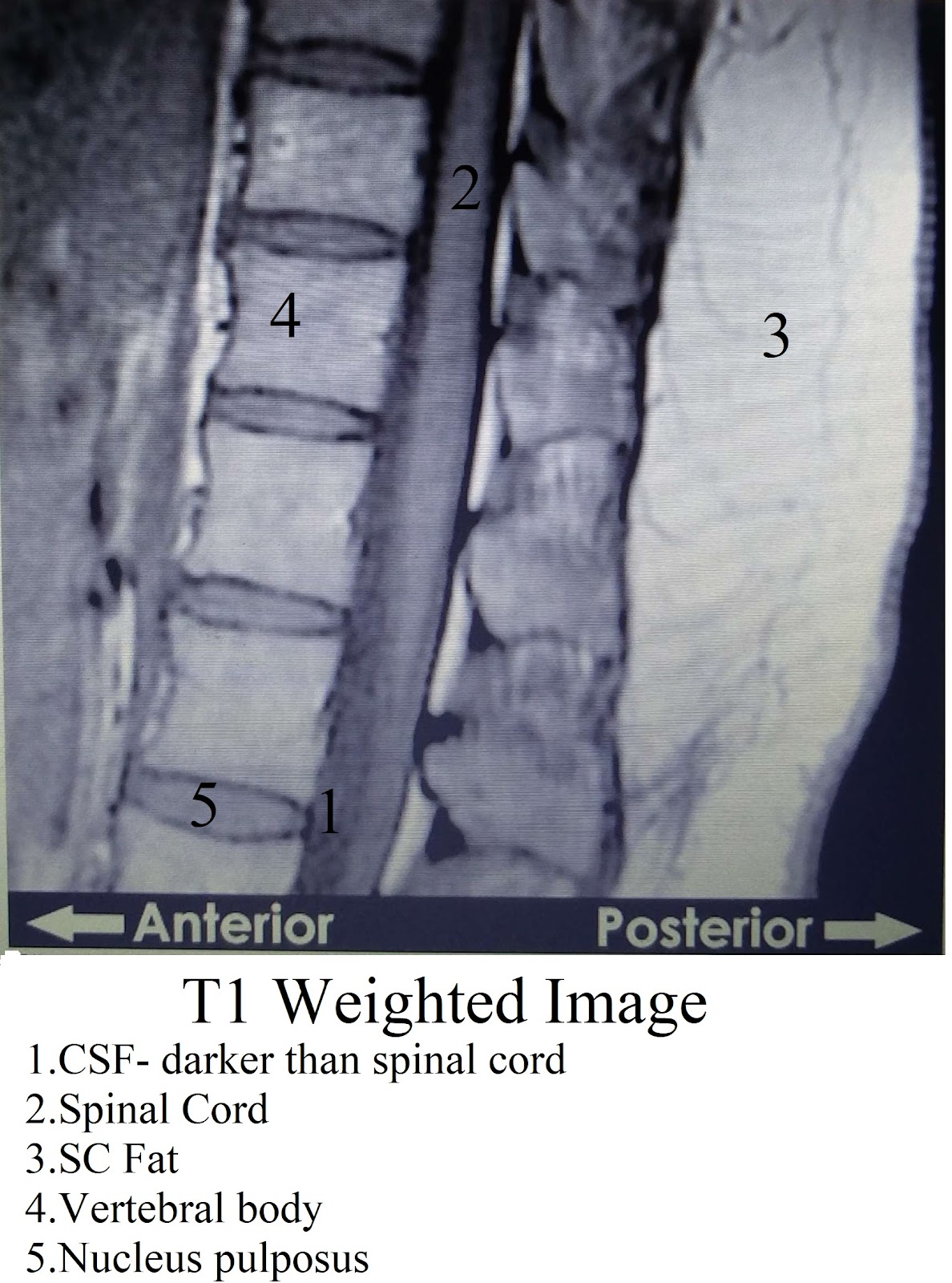
A: Compare it with the T1 image. Fat would appear white on both the images (subcutaneous fat in the above images) whereas water based tissue would appear dark on T1 and get highlighted on T2 images( CSF in the above photos).
The T1 weighted image is useful to delineate the normal anatomy whereas certain pathologies become easily appreciable with the T2 images.
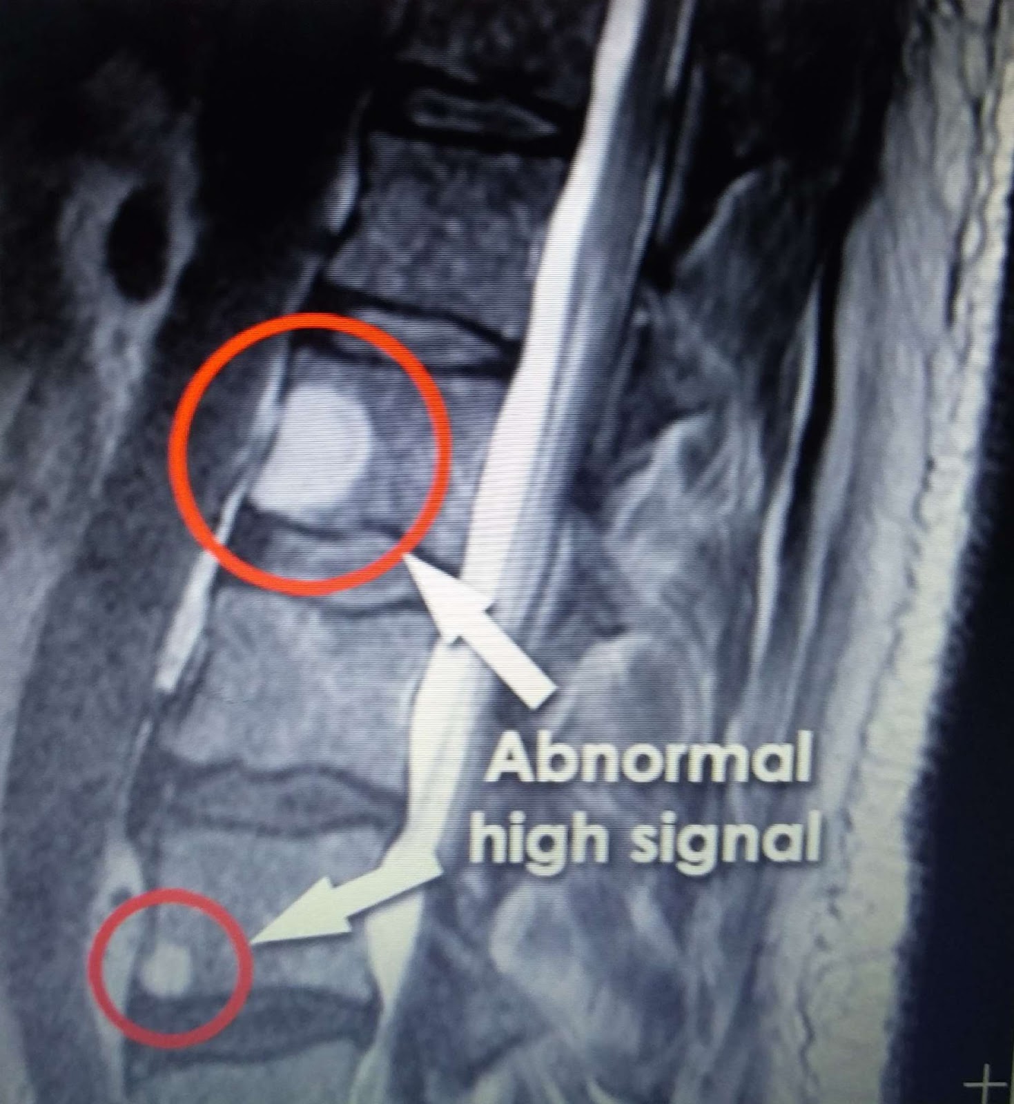
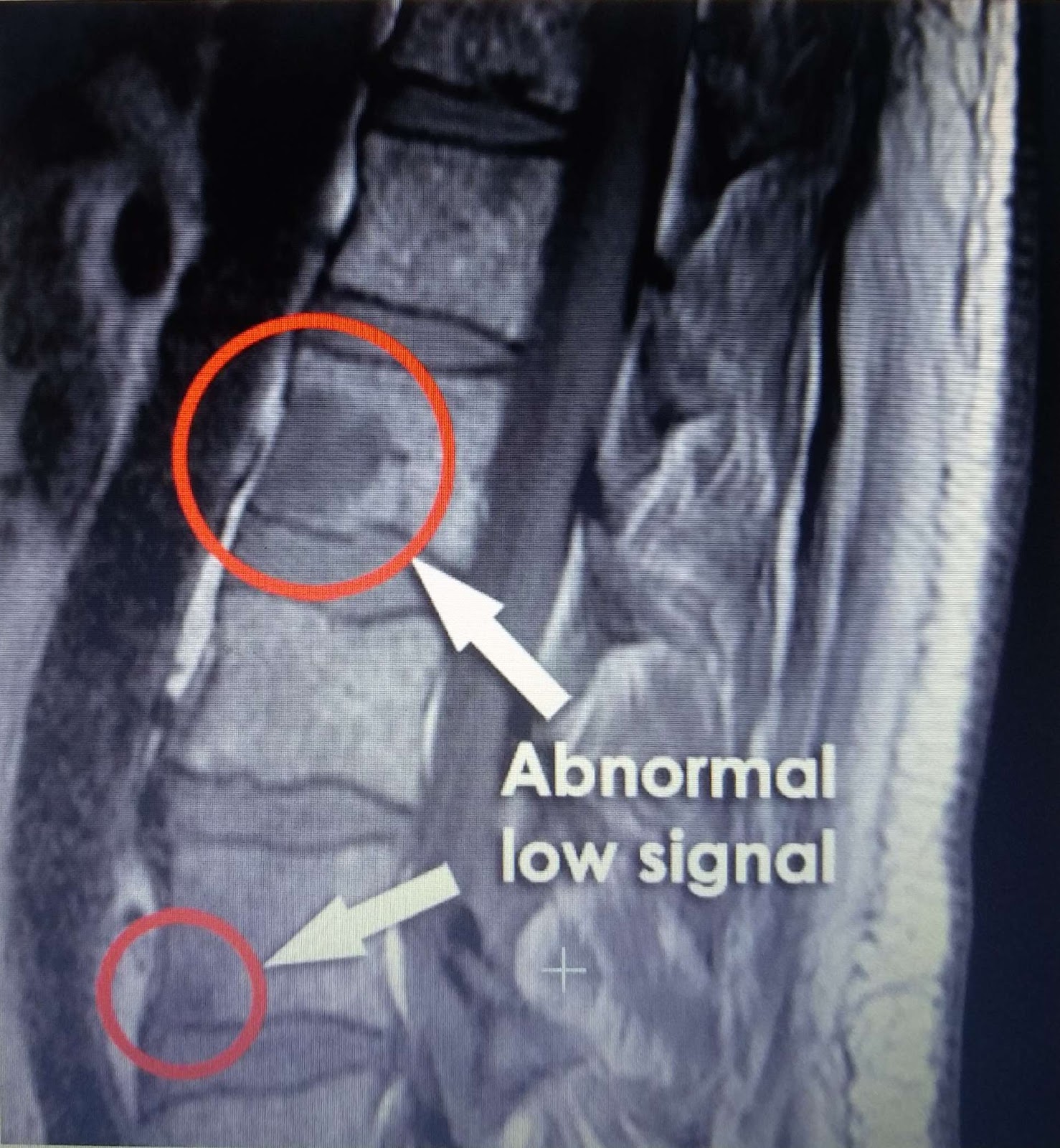
The above images are from a patient of multiple myeloma. The T2 image(above) helps to better appreciate the bony mets of the myeloma which understandably have higher water content and get highlighted.
Q: What is meant by T1 weighted and T2 weighted images?
Magnetic resonance imaging (MRI) makes use of the magnetic properties of protons which are present in water molecules, and therefore in all body tissues.
These protons are randomly oriented in the body.
The extremely powerful magnetic field of an MRI scanner aligns these protons along the field.
Radiofrequency pulses are subsequently applied which excite the protons.
The extremely powerful magnetic field of an MRI scanner aligns these protons along the field.
Radiofrequency pulses are subsequently applied which excite the protons.
Moments after the removal of the radiofrequency pulses the protons “relax” giving of their own radiofrequency signal.
This relaxing of the protons occurs by two methods: T1 Longitudinal relaxation ("spin-lattice") and T2 transverse relaxation( "spin-spin"/"slow dephasing")
During the process of T1 relaxation, protons reorient back to their longitudinal position in the magnetic field created. Fat quickly realigns its longitudinal magnetization and therefore appears bright on a T1 weighted image.
Conversely, water has much slower longitudinal magnetization realignment after an RF pulse and therefore, water has low signal and appears dark.
During the process of T2 relaxation, we look at something else: protons dephasing where spin becomes desynchronized.(Imagine a ball being spun and then returning to it's original position radially). Protons in water dephase slowly and this phenomenon is exploited in T2 weighted images.
and finally, a word about FLAIR:
Fluid Attenuated Inversion Recovery images in a way are inverse T2 images. The image from the free fluid, for example CSF, is suppressed instead of being highlighted. FLAIR images are particularly important to detect subtle changes at the periphery of the hemispheres and in the periventricular region close to CSF. One condition in which this is used commonly is Multiple sclerosis where periventricular white matter demyelination can be detected.

The area highlighted by the white arrow in the T2 image may be mistaken for CSF.The FLAIR image darkens the CSF and hence the remaining white hyperintense area is pathological.
Written by Pranav Hinduja


Comments
Post a Comment