Posts

Cherry on the Top

Image
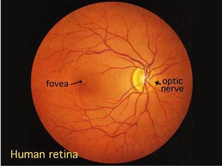
What is a Cherry Red Spot? A: To answer this we must start with the normal anatomy of the Macula : Macula Lutea (commonly called the yellow spot) is a circular area located at the centre of the posterior retina, 2 disc diameters from the optic disc. It has a slightly deeper red color when compared to the rest of the retina. The fovea is the central 1.5mm of the macula and has a higher density of cones and is responsible for color vision . A central depression in the fovea known as the foveola is completely devoid of ganglion cells and rods and has the highest density of cones and hence the most acute vision .It is also relatively thinner when compared to the rest of the retina. The Fovea receives its blood supply from the choroidal circulation via the long and short posterior ciliary arteries and NOT by the central retinal artery. Normal Retina Cherry Red Spot: A Cherry Red spot is a fundoscopic finding at the Macula in various diseases. It is basica...

Octreotide October

Image
Q: What is the basis of using octreotide is cases of Variceal Bleeding? A: Bleeding through esophageal varices is the major cause of death in patients with liver cirrhosis and portal hypertension. (1) According to the World Gastroenterology Organisation, the gold standard for treatment of acute variceal bleed is endoscopic band ligation and vasoactive pharmacological measures such as octreotide or terlipressin . These drugs can be given as adjunctive therapy, and also as primary therapy in primary centres where facilities for endoscopic evaluation and intervention may not be feasible. Other measures include, but are not limited to, sclerotherapy, portosystemic shunt surgeries, nonselective beta blockers and nitrates. In cirrhosis, portal hypertension is developed by a combination of: a) Increase of Intrahepatic resistance b) Increase of splanchnic blood flow, both contributing to the development of portal hypertension.(2) Treatment should thus be targeted at decreasing i...

MRIs: T1 & T2??

Image
T1 and T2 weighted images in MRI From the medical point of view the T1 and T2 weighted images are used to highlight different constituents of various tissues. T1 weighted images highlight only fat containing tissues in the body. T2 weighted images are timed to highlight both fat and water. One easy mnemonic to remember this: T1- 1 constituent is highlighted: Fat. T2- 2 highlighted constituents: Fat and Water. Note that the fat appears bright on both T1 and T2 images. Q:How do we differentiate fat from water based tissue on a T2 image since both are highlighted? A: Compare it with the T1 image. Fat would appear white on both the images (subcutaneous fat in the above images) whereas water based tissue would appear dark on T1 and get highlighted on T2 images( CSF in the above photos). The T1 weighted image is useful to delineate the normal anatomy whereas certain pathologies become easily appreciable with the T2 images. The above images are from a patie...

House-Surgeon and not House-Physician?

Image
Where the Term ‘House Surgeon’ Comes From Q: “Why is an intern called a house-surgeon and not a house-physician?” This is a very interesting question, and I had to dig really deep to find the answer. We’ve all heard this term being used for interns in India, but the matter was complicated by the term having different meanings in India and the UK. To understand where the term ‘house surgeon’ came from, we need to first understand who a ‘surgeon’ is. Who is a ‘surgeon’? The word ‘surgeon’ today refers to a doctor who specialises in the treatment of injuries or disorders of the body by incision or manipulation, especially with instruments. When you think of the word ‘surgeon’, I bet you think of someone like this: And not someone like this: Surprised? The English word ‘surgery’ comes from Latin ‘chirurgiae’, which in turn is derived from a Greek word meaning ‘the work of hand’. In medieval Europe, there used to be a huge divide between ‘physicians’ and ‘surgeo...

Cardiovascular Physics 101

Image
Q: WHY IS POISEUILLE’S EQUATION OR THE OHM'S LAW IMPORTANT IN THE CARDIOVASCULAR SYSTEM? A: Hemodynamics refers to the physical factors which govern the flow of blood. These physical factors are the same ones which dictate the flow of any fluid and are based upon the fundamental laws of physics. The laws of most relevance in the medical field are the Poiseuille’s equation and Ohm's Law. Poiseuille’s equation This relationship was first described in 19th century by the French physician Poiseuille and sheds light on the relationship between the primary determinants of resistance (R) to blood flow within a single vessel namely :- Vessel radius (r) Vessel length (L) Viscosity of blood (n) This tells us that resistance is directly proportional to both viscosity of blood and the length of the vessel and inversely proportional to the fourth power of the radius of the vessel. We can also infer that radius is the key determinant of the resistance to blo...

Running DMC: Diabetes, Metformin and Carbs.

Image
Type 2 Diabetes, Metformin & Carbs Q: “In the context of type 2 diabetes, metformin reduces the amount of sugar released into the bloodstream by the liver after a meal, but what happens to the unreleased sugar/carbs? Does it get converted to fat and stored or is passed out as feces?” A: Let’s begin with understanding the pathology in question and the mechanism of metformin’s action. What is the problem here? Diabetes (both types 1 and 2) turns the body into a predominantly catabolic state (as insulin is anabolic in nature). This means that for the cells of your body to get the fuel they need, the liver revs up synthesis of glucose from non-carbohydrate sources (amino acids, lipids), as glucose that has been absorbed from food is not entirely available for glycolysis and subsequent utilisation. Figure 1: Glycolysis - breakdown of available glucose into pyruvic acid, which can be utilised for cellular function. Source: https://www.checkdiabetes.org/glycolys...

Salmonella and Sickle Cell: Why?

Image
 Q: Why is Salmonella a common cause of joint infections in patients with sickle cell disease? Any particular reason? A: Salmonella is the most common cause of osteomyelitis (over 70%) in patients of sickle cell disease especially varieties like Salmonella enteritidis, Salmonella typhimurium, Salmonella paratyphi B, Salmonella choleraesuis and Salmonella aureus followed by Staphylococcus aureus , which is seen in 10% of patients suffering from sickle cell-related osteomyelitis. The majority of Salmonella infections in sickle cell patients involve bones (especially long bones) and joints and occur most frequently in early childhood. Multiple sites, often symmetrical, are usually involved. The following are the proposed/hypothesized reasons for the same: 1. At portal of entry: Capillary occlusion secondary to intravascular sickling may devitalize and infarct the gut, permitting Salmonella invasion. 2. In the blood: saturation of the macrophage system with red ...